
RESEARCH THEME
Quantification
We develop automated AI tools for measuring morphological and functional biomarkers.
OUR WORK IN QUANTIFICATION
Cardiac functional quantification from cine cardiac MR
Cardiologists routinely use cine cardiac MR scans to assess the health of the heart. Traditionally, deriving the functional metrics they need has been a laborious process, involving manually tracing round cardiac structures in multiple MR images. We have shown how AI-based segmentation tools can be used to automate this process, and furthermore implement automated quality control checks to ensure that the estimated metrics can be trusted by clinicians. Our metrics go beyond the commonly used metric of left ventricular ejection fraction, to quantify both diastolic and systolic function as well as the function of other cardiac chambers. This work has been commercially licensed to Perspectum who will use this technology in their COVERSCAN product for multi-organ health assessment.
Publications:
- "An AI Tool for Automated Analysis of Large-scale Unstructured Clinical Cine CMR Databases" European Heart Journal Digital Health, 2023. paper
- "Deep Learning for Classification and Selection of Cine CMR Images to Achieve Fully Automated Quality-Controlled CMR Analysis From Scanner to Report" Frontiers in Cardiovascular Medicine, 2021. paper
- "Fully Automated, Quality-Controlled Cardiac Analysis From CMR: Validation and Large-Scale Application to Characterize Cardiac Function" JACC: Cardiovascular Imaging, 2020. open access paper
- "Quality-aware Semi-supervised Learning for CMR Segmentation" Proceedings MICCAI STACOM, 2020. paper
- "Large-scale, Multi-vendor, Multi-protocol, Quality-controlled Analysis of Clinical Cine CMR Using Artificial Intelligence" European Heart Journal - Cardiovascular Imaging, Volume 22, Issue Supplement_2, 2021. abstract
Often, cardiac biomarkers are needed to monitor changes in function compared to those derived from a previous scan. Therefore, subsequent research has focused on the precision of the biomarker estimates rather than just their accuracy. We have proposed a method based on image interpolation of the short axis stack and also evaluated a deep learning based tool for predicting dense 3D segmentations from sparse slices. Both methods were able to narrow Bland-Altman scan-rescan confidence intervals for all biomarkers.
Publications:
- "Improving the Scan-rescan Precision of AI-based CMR Biomarker Estimation" Proceedings MICCAI STACOM, 2024. paper
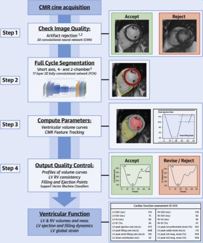
Tissue quantification from cardiac MR T1 imaging
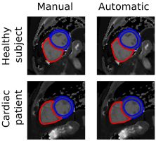
We also developed a similar pipeline for automated quantification of ShMOLLI T1 data. This pipeline also featured robust quality control steps, this time based upon segmentation uncertainty. Disease-specific reference ranges from the UK Biobank dataset were published for the first time.
Publications:
- "Automated Quantification of Myocardial Tissue Characteristics From Native T1 Mapping Using Neural Networks With Uncertainty-based Quality-control" Journal of Cardiovascular Magnetic Resonance, 22:60, 2020. open access paper
Cardiac functional quantification from echocardiography
Although cardiac MR is considered to be the "gold standard" imaging modality for assessment of most aspects of cardiac function, by far the most commonly used modality is cardiac ultrasound, or echocardiography. Most work on AI-based functional assessment from echocardiography has focused on ventricular volumes at just the end-diastole and end-systole phases, which can then be used to calculate the ejection fraction. We have shown how AI can be used to estimate a wider range of functional biomarkers and compared the results to those estimated from cardiac MR.
Publications:
- "AI-enabled Assessment of Cardiac Systolic and Diastolic Function from Echocardiography" Proceedings MICCAI ASMUS, 2022. paper
Spleen size assessment from ultrasound
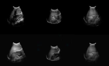
Assessment of the size of the spleen is a crucial task in the monitoring of patients with Sickle Cell Disease (SCD). Enlargement of the spleen in SCD patients, known as splenomegaly, can have associated health complications and even be life-threatening. The standard clinical workflow for spleen size assessment involves manual length measurement from 2D ultrasound, but this is crude and does not provide an estimate of volume, which is potentially a much better biomarker of spleen size. In this work, we developed a deep learning-based framework for automatically estimating spleen volume from routinely-acquired 2D ultrasound images. The method was trained using a dataset of highly realistic synthetic ultrasound images derived from CT scans using a semantic ultrasound diffusion model, which we made publicly available.
Publications:
- "DeepSPV: A Deep Learning Pipeline for 3D Spleen Volume Estimation From 2D Ultrasound Images" Medical Image Analysis, 2025. open access paper
Dataset:
Dataset of realistic synthetic spleen ultrasound images with ground truth volume data
Interested in our work?
Explore our research or get in touch with the group.
Contact the group →